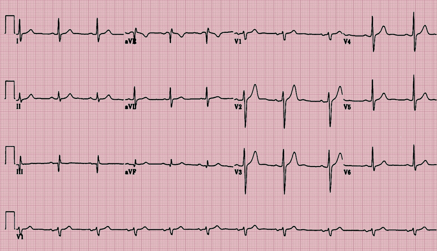
Echocardiography or Electrocardiograms (ECG)
Echocardiography or Electrocardiograms — also called ECGs. An electrocardiogram (ECG) records the electrical sign from your heart to check for various heart conditions. Electrodes are placed on your chest to record your heart’s electrical signals, which causes your heart to beat. The signs are shown as waves on a connected computer monitor or printer.
Dr.Shriniwas Mante who have special expertise and exhaustive in-depth experience of Sonologist and Radiologist.
You may require an ECG if you have any of the following signs and symptoms:
- Chest pain
- Heart palpitations
- Fast heartbeat
- Shortness of breath
- Weakness, or a decrease in the capacity to work out.
How the Test is Performed?
You will be asked to lie down. The health care provider will clean several areas on your arms, legs, and chest, and then will attach small patches called electrodes to those areas. It may be necessary to shave or clip some hair so the patches stick to the skin. The number of patches used may vary.
The patches are connected by wires to a machine that turns the heart's electrical signals into wavy lines, which are often printed on paper. The doctor reviews the test results.
You will need to remain still during the procedure. The provider may also ask you to hold your breath for a few seconds as the test is being done.
It is important to be relaxed and warm during an ECG recording because any movement, including shivering, can alter the results.
Sometimes this test is done while you are exercising or under light stress to look for changes in the heart. This type of ECG is often called a stress test.

