2D ECHO
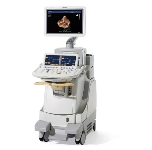
2D Echocardiography or 2D Echo of heart is a test in which ultrasound technique is used to take pictures of heart. It will be displayed in a cross-sectional ‘slice’ of the beating heart, showing chambers, valves and the major blood vessels of heart.
Dr.Shriniwas Mante who have special expertise and exhaustive in-depth experience of Sonologist and Radiologist.
It’s a non invasive heart evaluation that makes pictures of the segments of the heart with sound vibrations.It represents the different parts of the heart like in pictures in order that it becomes simple to check if there’s a harm or blockages, and flow of blood speed.
2D Echocardiography or 2D Echo as it’s commonly known as, is a test used to diagnose coronary disorders. This test also can help to determine the quantity and speed of blood circulation through each of the chambers of heart disease.
How is 2D Echo done?
Patient is made to change in a front open robe and a colourless gel is applied to the chest area. Then he is asked to lay on his left side as the technician moves the transducer across the various parts of his chest to get specific/desired views of the heart. Instructions may also be given to the patient to breathe slowly or to hold it. This helps in getting superior quality pictures. The images are viewed on the monitor and recorded on paper, video or DVD. The cardiologist later reviews and interprets the recordings.
What it detects?
Echocardiography is a significant tool in providing the physician important information about heart on the following:
![]() Size of the chambers, volume and the thickness of the walls.
Size of the chambers, volume and the thickness of the walls.
![]() Pumping function, if it is normal or reduced to a mild/severe degree.
Pumping function, if it is normal or reduced to a mild/severe degree.
![]() Valve function – structure, thickness, and movement of heart’s valves.
Valve function – structure, thickness, and movement of heart’s valves.
![]() Volume status as low blood pressure may occur as a result of poor heart function.
Volume status as low blood pressure may occur as a result of poor heart function.
![]() Pericardial effusion (fluid in the pericardium and the sac that surrounds the heart), congenital heart disease, blood clots or tumors, abnormal elevation of pressure within the lungs etc.
Pericardial effusion (fluid in the pericardium and the sac that surrounds the heart), congenital heart disease, blood clots or tumors, abnormal elevation of pressure within the lungs etc.
How long does 2D Echocardiography take?
A brief exam in normal case may be done within 15-20 minutes. However, when there are heart problems, it may take much longer.
How safe is echocardiography?
It is absolutely safe. There are no known risks of the ultrasound in this type of testing. To live healthy & happy, one must keep a check on the body’s functioning by going for regular health checkups. This helps in assessing risk factors and also for diagnosing diseases at an early stage, which will result in effective treatment and better management of the condition.


